Page Not Found
Page not found. Your pixels are in another canvas.
A list of all the posts and pages found on the site. For you robots out there is an XML version available for digesting as well.
Page not found. Your pixels are in another canvas.
About me
This is a page not in th emain menu
Hey everyone! Thanks for visiting my website; I am currently in the process of building the site structure, adding files/images, and writing content for subpages. I added the blog section in here because this was built in from the “academicpages” theme that I forked from Github… We will see how many blog posts I actually make…
Roberts, G. S., Young, S. M., Lancaster, J., Siegel, S. F., and Caruso, A. N. (2017). "Implementing a Low-Cost Sawyer-Tower Circuit to Measure Ferroelectric Properties at Various Frequencies" [Unpublished bachelor's thesis]. University of Missouri - Kansas City.
Roberts, G. S., Francois, C. J., Starekova, J., Roldan-Alzate, A., and Wieben, O. (2022). "Non-invasive assessment of mesenteric hemodynamics in patients with suspected chronic mesenteric ischemia using 4D flow MRI". Abdom Radiol, 47(5), 1684-1698. doi:10.1007/s00261-020-02900-00
Macdonald, J. A., Roberts, G. S., Corrado, P. A., Beshish, A. G., Haraldsdottir, K., Barton, G. P., . . . Wieben, O. (2021). "Exercise-induced irregular right heart flow dynamics in adolescents and young adults born preterm". J Cardiovasc Magn Reson, 23(1), 116. doi:10.1186/s12968-021-00816-2
Oechtering, T. H., Roberts, G. S., Panagiotopoulos, N., Wieben, O., Roldan-Alzate, A., and Reeder, S. B. (2021). "Abdominal applications of quantitative 4D flow MRI". Abdom Radiol. doi:10.1007/s00261-021-03352-ww
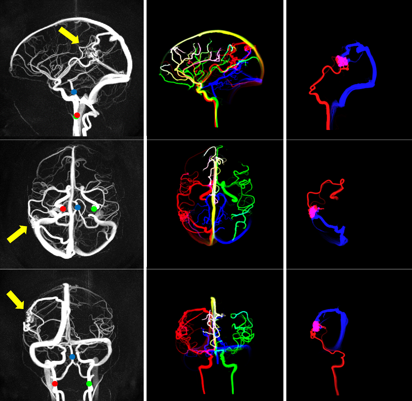
Roberts, G. S.*, Loecher, M. W.*, Spahic, A., Johnson, K. M., Turski, P. A., Eisenmenger, L. B., and Wieben, O. (2022). "Virtual injections using 4D flow MRI with displacement corrections and constrained probabilistic streamlines". Magn Reson Med, 87(5), 2495-2511. doi:https://doi.org/10.1002/mrm.29134
*Authors contributed equally to this work.
Oechtering, T. H., Roberts, G. S., Panagiotopoulos, N., Wieben, O., Reeder, S. B., and Roldan-Alzate, A. (2022). "Clinical Applications of 4D Flow MRI in the Portal Venous System". Magn Reson Med Sci, 21(2), 340-353. doi:10.2463/mrms.rev.2021-0105d
Roberts, G. S., Peret, A., Koscik, R. L., Jonaitis, E. M., Hoffman, C. A., Rivera-Rivera, L. A., Cody, K. A., Rowley, H. A., Johnson, S. C., Wieben, O., Johnson, K. M., & Eisenmenger, L. B (2023). "Normative Cerebral Hemodynamics in Middle-Aged and Older Adults using 4D Flow MRI: Initial Analysis of Vascular Aging". Radiology. 307(3):e222685. doi: 10.1148/radiol.222685
Roberts, G. S.*, Hoffman, C. A.*, Rivera-Rivera, L. A., Berman, S. E., Eisenmenger, L. B., & Wieben, O. (2023). "Automated Hemodynamic Assessment for Cranial 4D Flow MRI". Magnetic Resonance Imaging, 97:46-55. https://doi.org/10.1016/j.mri.2022.12.016
*Authors contributed equally to this work.
Eisenmenger, L. B.*, Peret, A.*, Famakin, B. M., Spahic, A., Roberts, G. S., Bockholt, H. J., Johnson, K. M., & Paulsen, J. S. (2023). "Vascular Contributions to Alzheimer’s Disease". Translation Research, 254:41-53, https://doi.org/10.1016/j.trsl.2022.12.003.
*Authors contributed equally to this work.
Huang, A., Roberts, G. S., Reeder, S. B., & Oechtering, T. H. (2023). "Reference Values for 4D Flow Magnetic Resonance Imaging of the Portal Venous System". Abdominal Radiology, 48(6):2049-2059. doi: 10.1007/s00261-023-03892-3
Capel, K. W., Roberts, G. S., Kuner, A. D., Manunga, J., Chang, W., Spahic, A., Peret, A., Wieben, O., Johnson, K. M., & Eisenmenger, L. B. "Beyond Time-of-Flight MRA - Review of Flow Imaging Techniques". Accepted to Neurographics.
Capel, K. W., Roberts, G. S., Kuner, A. D., Manunga, J., Chang, W., Spahic, A., Peret, A., Wieben, O., Johnson, K. M., & Eisenmenger, L. B. Beyond Time-of-Flight MRA: Review of Flow Imaging Techniques. Accepted to Neurographics.
Carter, K. J., Ward, A. T., Kellawan, J. M., Harrell, J. W., Peltonen, G. L., Roberts, G. S., Al-Subu, A., Hagen, S. A., Serlin, R. C., Eldridge, M., Wieben, O., & Schrage, W. G. (2023). "Reduced basal macrovascular and microvascular cerebral blood flow in young adults with metabolic syndrome: potential mechanisms". Journal of Applied Physiology. doi: 10.1152/japplphysiol.00688.2022
Eisenmenger, L. B.*, Peret, A.*, Roberts, G. S., Spahic, A., Tang, C., Kuner, A., Grayev, A., Field, A., Rowley, H. A., & Kennedy, T. (2023). "When Less is More: FAST MR Protocols for Neuroradiology". Radiographics. doi: 10.1148/rg.220147
*Authors contributed equally to this work.
Annual UW-Madison MRI Group meeting to share findings or discuss relevant MRI topics. In this talk, I discuss the findings from our chronic mesenteric ischemia study. After eating a meal, blood flow to the gut normally increases. In patients with chronic mesenteric ischemia (CMI), this blood flow response is stunted, which can be caused by atherosclerotic narrowing, arteritis, or other pathologies that restrict blood flow. In this work, we use 4D flow MRI to measure mesenteric blood flow before and after a meal and show that blood flow is stunted in patients with CMI relative to controls, making 4D flow MRI a potential diagnostic tool.
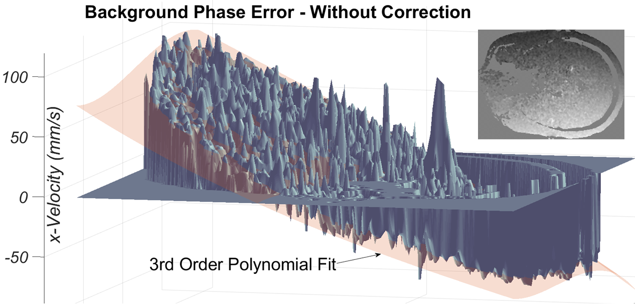
My first in-person presentation was a powerpitch at SMRA in 2019. Powerpitches are typically short (~3 minute) elevator pitches designed to quickly grab the interest of the audience. For this work, I discuss the findings from our study on the effects of automating background phase correction in our reconstruction pipeline. When we were initially developing our cranial 4D flow analysis tool, we were constantly looking for ways to decrease manual interaction and the time needed to load 4D flow data for post-processing. One of the manual steps in our processing pipeline was background phase correction, which corrects eddy-current phase errors created by the flow-encoding gradients. In this presentation, I showed that an automatic phase correction implementation in the reconstruction actually outperforms manual background phase correction. This justified the use of skipping the manual correction step if background phase correction was done in the reconstruction, streamlining our tool.
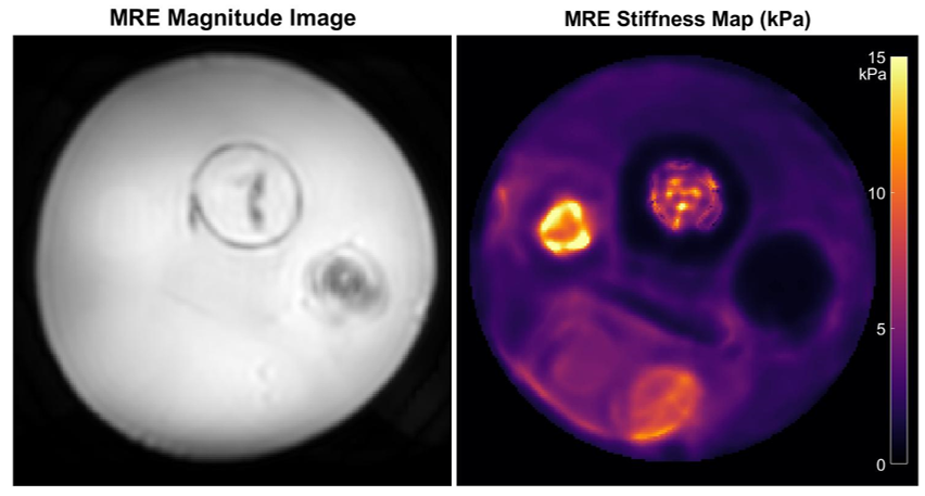
Annual UW-Madison MRI Group meeting to share findings or discuss relevant MRI topics. For this talk, I reviewed brain MR elastography (MRE), a technique that allows one to measure brain stiffness using MRI (crazy!). We recently collaborated with GE Healthcare and the Mayo Clinic to implement brain MRE at our institution. This talk was designed to provide background on our recently-implemented MRE technique for our MRI colleagues.
This talk was the first of a 2 part series, intended for members of the Wisconsin Alzheimer’s Disease Research Center (ADRC) to demonstrate our state-of-the-art vascular imaging techniques that our research groups have developed for studying cerebrovascular contributions to dementia and Alzheimer’s disease. Specifically, we focus on basic principles of flow MR imaging, what can be measured and how we measure it, and some results of recent studies.
This was the second talk of our 2-part ADRC vascular imaging series. In the previous lecture, we discussed principles of flow imaging and results from recent studies. In this lecture, we focus on more advanced imaging methods to obtain non-conventional measures of: vessel stiffness, brain tissue stiffness (MRE), perfusion (ASL), and ways to assess glymphatics and the integrity of the blood brain barrier.
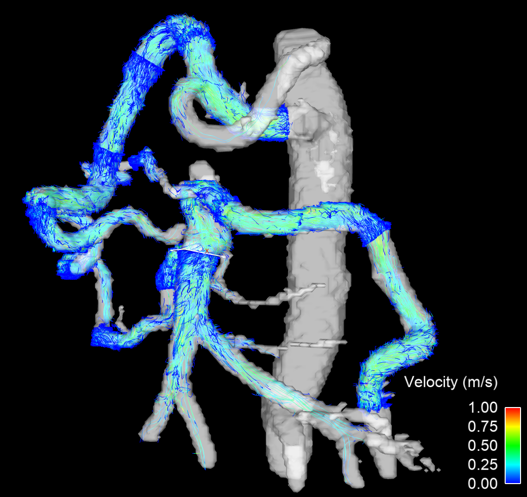
I presented an abstract as a second author (originally submitted by my advisor Laura Eisenmenger) on the use of 4D flow-generated virtual injections for use in venous mapping of arteriovenous malformations (AVM). AVMs range from benign to very high risk, depending on the size, location, number of feeding vessels, etc. Because of this, they can difficult to manage and treat. Transvenous embolization has recently emerged as a promising surgical approach to treat AVMs. In this presentation, we discuss the promise of our virtual injection technique (using forward/reverse injections) in an AVM case at our institution.
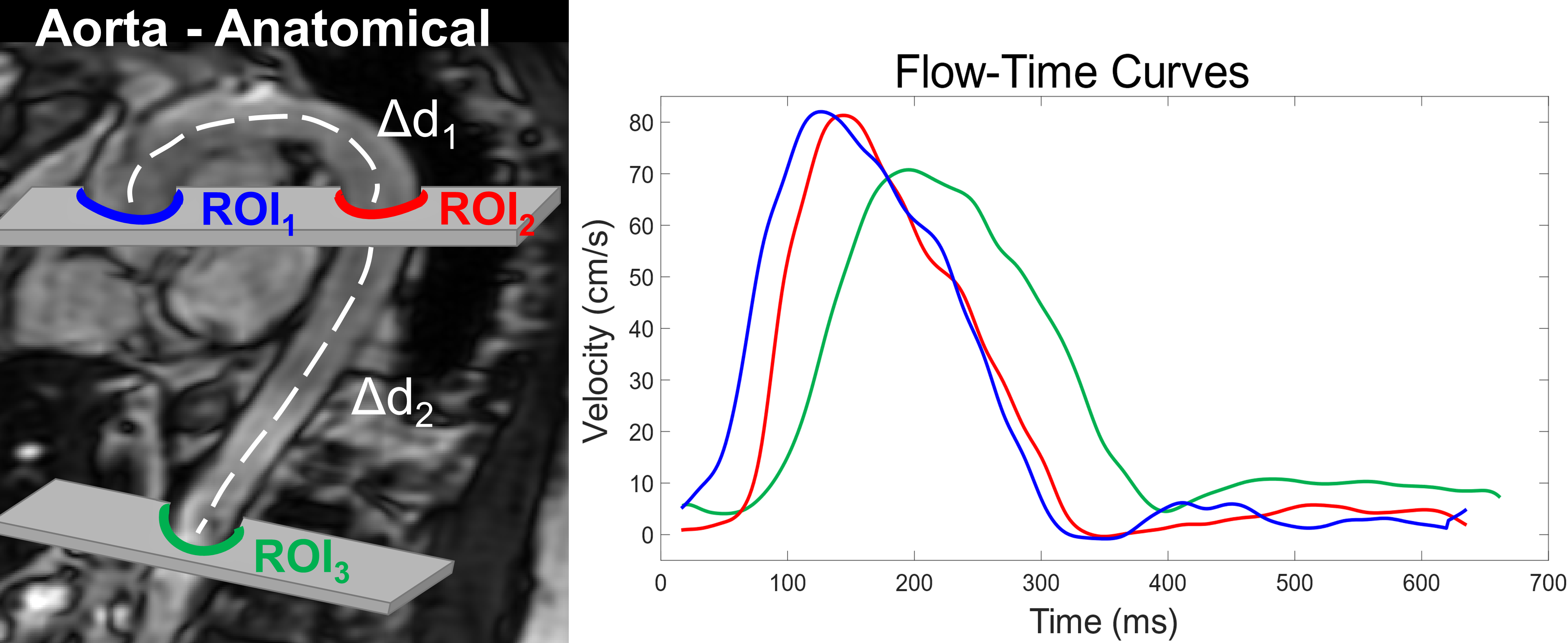
In this oral presentation given at SMRA, I discuss our findings from our free-breathing pulse wave velocity (PWV) study in collaboration with the Okonkwo lab. Here, we assessed the feasibility of using radial 2D phase contrast MRI to measure aortic PWV, which is directly related to aortic stiffness. Because respiratory motion during a scan can result in bad image quality, PWV MRI scans are often performed under breath-hold conditions which can be sometimes be challenging. For this project, we allowed subjects to breath freely during the scan and used respiratory gating to retrospectively create images only in stationary respiratory phases, improving image quality. We also used regularized (local low rank) reconstructions to greatly improve image quality. We demonstrated no significant differences between our free-breathing technique and the standard breath-hold technique.
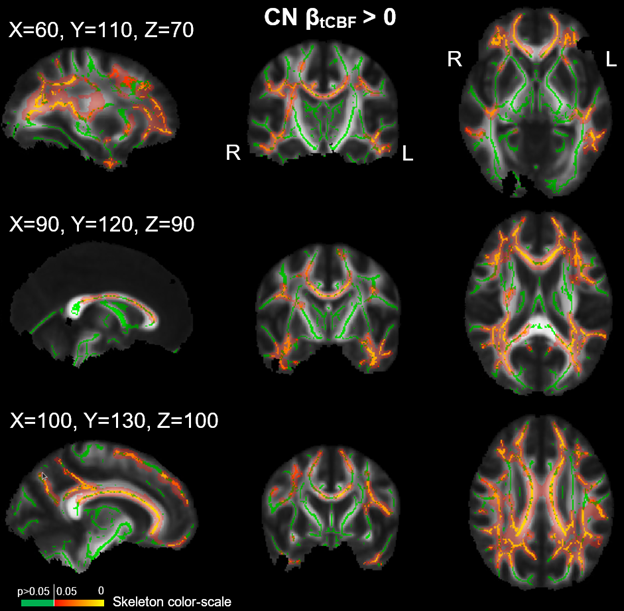
This oral presentation was given at ISMRM on the work related to the (ongoing) NODDI - 4D Flow study. In this talk, I focus on the relationship between macrovascular flow and pulsatility and observed white matter microstructure in normal subjects and Alzheimer’s disease subjects.
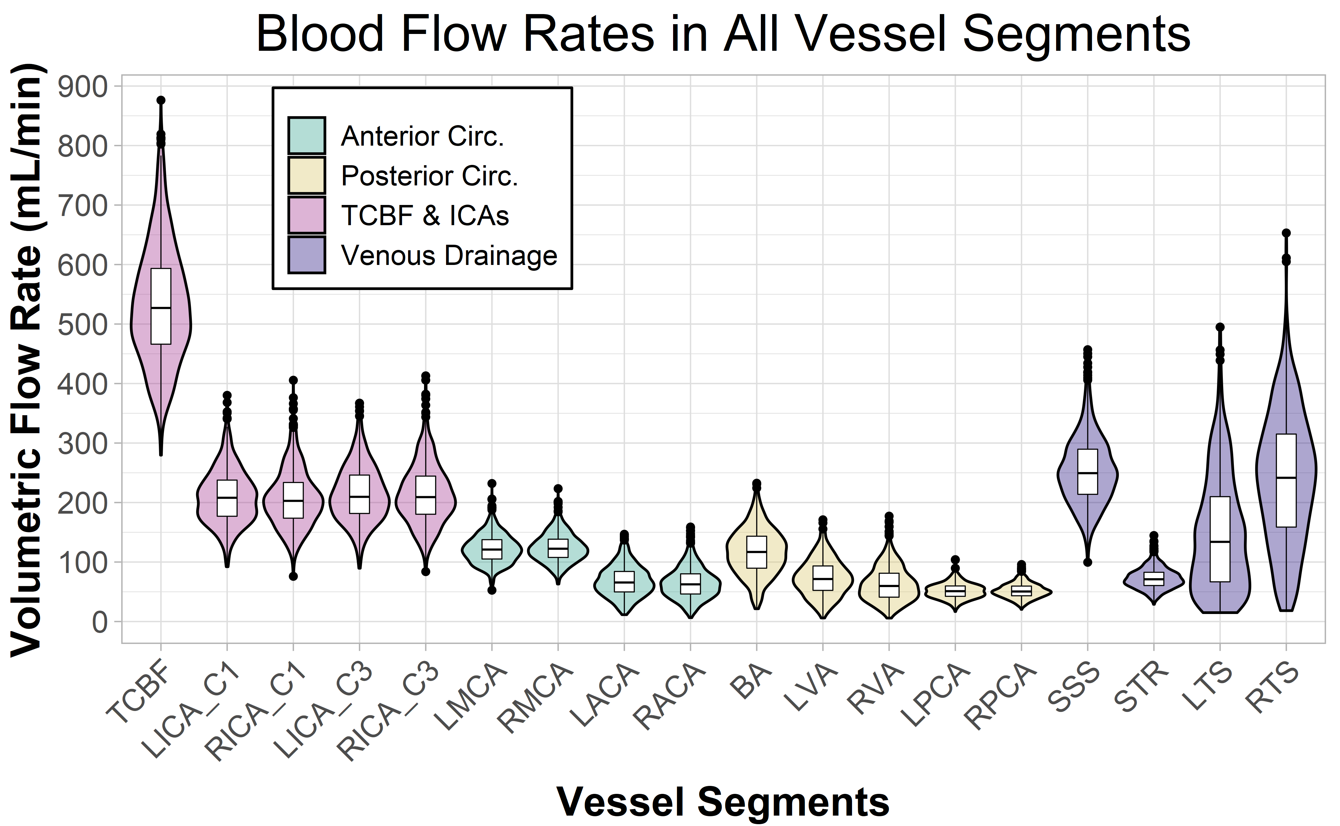
This was our first oral presentation on the work that Anthony Peret and I have done on the normative 4D flow study. This talk focused on methodology and basic statistics, specifically our custom cranial 4D flow analysis tool, intra/interobserver repeatability, normal intracranial blood flow ranges in older adults, and how blood flow and pulsatility change as we age. It was presented at ISMRM and was my first in-person oral presentation since COVID. Anthony is currently preparing a similar talk for late August 2022 at ASFNR on the relationship between vascular risk factors and measured intracranial flow and pulsatility.
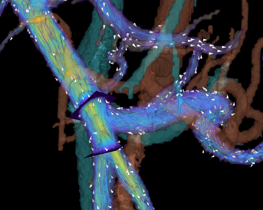
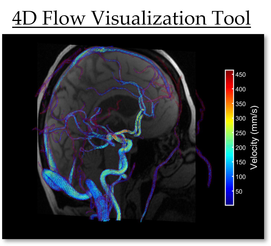
4D flow MRI (time-resolved, volumetric, three-directional velocity-encoded MRI) is a powerful imaging method that can capture blood flow, vascular anatomy, and complex flow patterns in a single acquisition.
Phase-contrast magnetic resonance imaging (PC-MRI) is a non-invasive imaging method capable of quantifying time-resolved hemodynamics. This dissertation aims to develop novel acquisition methods and post-processing tools for the robust assessment of intracranial and aortic hemodynamics using PC-MRI and to study their relationship with aging and neurodegeneration.